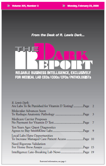
Molecular Advances Soon To Reshape Anatomic Path

Predictions that pathology is to become more quantitative because of new technologies

CEO SUMMARY: Early this month, the second annual Molecular Summit assembled molecular first movers and early adopters to discuss their efforts to integrate molecular imaging and molecular diagnostics in patient care. One clear message emerged from two days of presentations and discussion: a host of new technologies is ready for clinical introduction and is likely to rapidly transform both radiology and pathology.

HEADING TO AN ANATOMIC PATHOLOGY GROUP near you is a flood of molecular technologies that promise to swiftly transform the pathology profession as we know it today.

That was a common view of both speakers and attendees at this year’s Molecular Summit on Integration of In Vivo and In Vitro Diagnostics, which took place earlier this month in Philadelphia. It is not often that such a broad consensus emerges from an event like this, which adds impact to this development.

The consensus centered around these key points:

• Many molecular technologies in both imaging and in vitro diagnostics presented at Molecular Summit are almost ready for clinical use and will be introduced on an unexpectedly short timeline.

• Many of these molecular technologies will give pathologists and radiologists remarkably powerful and precise new capabilities in the diagnosis of disease.

• Personalized medicine has arrived in healthcare and new personalized medicine services are being introduced into our healthcare system at a steady pace.

• Diagnostics will increasingly be organized around the use of quantitative data, particularly in anatomic pathology.

• Single-analyte assays will give way to multi-analyte assays. These new tests will often incorporate tens of thousands of analytes and data points for evaluation.

• Multi-modality analysis will become quite common and will utilize molecular imaging, molecular diagnostics, and other types of clinical data for assessment in diagnosis, therapeutic decisions, and monitoring patient progress.

Molecular Summit is organized by THE DARK REPORT to bring together first movers and early adopters in pathology and radiology who are developing integrated clinical services. It is unique in the world because it is the only conference which brings together experts in molecular imaging, molecular diagnostics, and health informatics.

Several speakers predicted that the natural evolution of genetic medicine and molecular technologies will encourage the creation of a single diagnostic service, particularly in academic and tertiary care centers. This single diagnostic service will be capable of providing clinicians with an integrated diagnosis. It will also offer clinicians active consultation with the pathologists and radiologists who evaluated the specimens and worked up the patient.

Just such an integrated diagnostic service is under development at the UCLA Medical Center in Los Angeles, California. The departments of pathology and radiology are preparing to open an integrated diagnostics center. Jonathan Braun, M.D., Ph.D., Chair, UCLA Department of Pathology and Laboratory Medicine, described the project to the Molecular Summit attendees. Dieter Enzmann, M.D., Chair of the UCLA Department of Radiology, was in attendance and is a collaborator on this integrated diagnostic service.

Patient-Centered Care

Braun described the effort as patient-centric care delivered by an integrated radiology/pathology service model. The two departments will build their first integrated radiology/pathology patient service center in a physician office building in Santa Monica. They expect the promise of an integrated diagnosis within 48 to 72 hours will encourage local physicians to refer patients—thus expanding market share for both the UCLA radiology and pathology departments.

Their goal is to deliver a novel, patient-friendly approach to image-guided biopsy sampling for malignancy work-up. Radiologists will use CAT, MRI, and ultra-sound to isolate and sample suspicious lesions. Pathologists will handle processing and diagnosis of the specimen. The pathology report will be combined with the radiology report to provide referring physicians with a comprehensive single-copy report that is accessible via the Internet.

For anatomic pathologists, one unmistakable theme delivered by multiple speakers was that a host of new technologies is about to transform the profession. Speaker Richard C. Friedberg, M.D., Ph.D., Professor and Deputy Chairman Department of Pathology at Tufts University School of Medicine, in Springfield, Massachusetts, described how oncology is rapidly adopting a molecular classification of different cancers. He noted that this has already happened in hematologic oncology.

Quantitative Diagnosis

“The trend in diagnostics is clearly towards integration of data,” stated Friedberg. “Anatomic pathology and radiology have traditionally been qualitative ‘pattern recognition’ fields. However, new technologies are providing practitioners in both specialties with the greater precision, accuracy, reliability, and measurability needed to become more quantitative.”

Friedberg then noted that clinical pathology has long been a quantitive diagnostic service, stating “clinical pathology regularly gathers information from a number of sources within and without the laboratory and delivers an integrated answer to the physician. In this same manner, anatomic pathology is going to evolve into primarily a quantitive service.”

Computer-Aided Diagnosis

It is likely that both computer-aided diagnosis (CAD) and pattern recognition technologies will play an important role in anatomic pathology’s transition from a qualitative service to a quantitative service. Several speakers are doing advanced work in these fields and demonstrated these techniques.

Based on his work with prostate and breast cancer, Anant Madabhushi, Ph.D., Assistant Professor, Director, Laboratory for Computational Image & Bio-infomatics (LCIB) at Rutgers The State University of New Jersey in Parsippany, showed how multi-modality diagnosis can be achieved using enhanced informatics and computer-aided image diagnosis.

Madabhushi showed ways that CAD can be used to evaluate MRI images. CAD can characterize MRI data using multiple texture features. Unsupervised consensus clustering is also used to classify data. Madabhushi noted that high sensitivity and specificity (>90%) can be achieved on a per-voxel basis.

Next, Madabhushi showed the use of CAD with the histological images. For a prostate cancer case, he demonstrated how the CAD software can isolate regions of interest for the pathologist’s examination. CAD will also identify different regions for evaluation to determine the grading scheme of the cancer.

Pattern Recognition

Pattern recognition will be another transformational technology in anatomic pathology. It figures prominently in the work of Badri Roysam, Ph.D., Professor of Electrical Computer & Systems Engineering at Rensselaer Polytechnic Institute in New York City.

In radiology, Roysam described the need for more biochemically-specific information at the cellular scale. “This need spurred the development of high throughput gene and protein analysis tools, such as microarrays,” noted Roysam. “But these tools miss all spatially-linked information. We want to understand the intact tissue structure, the spatial location of markers, and the spatial relationships among multiple cells and tissue. Only imaging can provide these forms of information.”

In histopathology, Roysam described how the field is evolving along these lines:

• “Multi-scale Multiplex Histocytometry”, which means: 1) understanding complex tissues on a tissue scale with sub-cellular detail; 2) being able to see all the molecules of interest in their native context; and 3) being able to quantify at multiple scales.

• Optical microscopy will remain an attractive tool for molecular histopathology, with these points: 1) growing role for fluorescent stains alongside chromogenic stains; 2) higher level of multiplexing will allow us to examine many molecules of interest in their context (many more than humanly viewable in brightfield); and 3) high-throughput (arrays) & high-extent (whole-slide) imaging.

• Growing role for automated image scoring and interpretation that produces more quantitative and objective inferences, along with data-based scores that are consistent across [testing] centers.

These highlights from Molecular Summit 2009 demonstrate why first movers in pathology and radiology are making swift progress toward the ideal of an integrated diagnostic service. At the same time, new technologies are arriving that promise to accelerate this evolution.

George Poste Predicts Major Role for Dx

KEYNOTE SPEAKER George Poste, DVM, Ph.D., discussed the likely adoption paths for molecular diagnostics and molecular imaging. He is Chief Scientist and Director at The Biodesign Institute of Arizona State University, located in Tempe, Arizona.

Poste is one of the world’s foremost experts on biomarkers and was bullish on the role that molecular diagnostics will play in realizing the goals of personalized medicine. He outlined how healthcare will utilize diagnostics as it evolves and discussed the bullet points below:

Central Role of Next-Generation Diagnostic Technologies in Proficient Healthcare Delivery

  • Precision diagnosis
  • Rational Rx selection
  • Reduce errors
  • Increasingly standardized clinical practice
  • Remote health status monitoring and patient compliance
  • Disease predisposition and risk mitigation
  • Increased personal responsibility for risk mitigation and wellness
  • Integrated care continuum


David Galas Discusses Systems Biology

INTRODUCING MANY IN THE AUDIENCE to developments in the field of systems biology was David Galas, Ph.D. He is Professor at the Institute for Systems Biology, in Seattle, Washington, and Vice President and Chief Scientific Officer of the Battelle Memorial Institute in Columbus, Ohio.

Galas described how systems biology is an approach to dealing with the complexity of human biology. He discussed how biological networks capture, transmit, process, and channel information. His team is working to develop multi-analyte diagnostic capabilities using this information, as follows:

Technologies Key to Catalyze Future Personalized Medicine

  • High throughput DNA sequencing for individual human genome sequences
  • Targeted MRM protein mass spectrometry for discovery and validation of blood protein diagnostic fingerprints
  • Microfluidic protein and RNA chips to measure blood organ-specific protein, and RNA fingerprints and to type millions of individuals
  • New chemistry and new approaches to protein-capture agents
  • Single-cell and single-protein analyses—deciphering the interplay of the digital genome and the environment
  • In vivo and in vitro molecular imaging to detect, assess disease distribution and follow therapy


Cliff Hoyt Explores Future of Tissue Sections

ANATOMIC PATHOLOGISTS WERE CAREFUL LISTENERS during the presentation of Cliff Hoyt, M.S., Vice President and Chief Technology Officer at CRI-Inc. in Woburn, Massachusetts.

Hoyt’s company is using technologies in informatics and pattern recognition to develop new capabilities in in vitro diagnostics. He predicted that molecular technologies will use tissue in different ways, as noted below:

Watch For these Tissue Section-based Molecular Technologies

  • Methods that avoid paraffin embedding
  • Methods of fixation that preserve labile proteins (e.g., phospho-proteins)
  • Biopsy methods

– Needle vs resection

– Cryo-needles

  • Circulating tumor-based molecular profiling (blood and urine)
  • Imaging mass spectroscopy

Comments

Leave a Reply

;

You are reading premium content from The Dark Report, your primary resource for running an efficient and profitable laboratory.

Get Unlimited Access to The Dark Report absolutely FREE!

You have read 0 of 1 of your complimentary articles this month

Privacy Policy: We will never share your personal information.